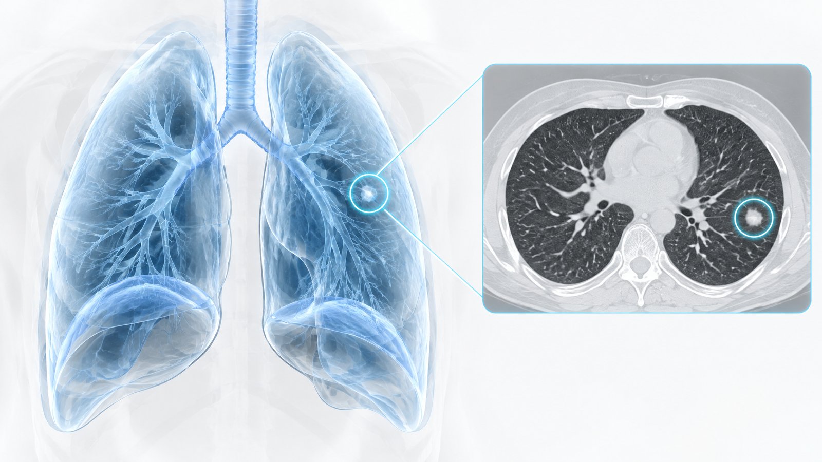
Four months after founding, our small team has reached our first technical milestone: a working proof-of-concept model that detects small pulmonary nodules on chest CT scans.
Why lung nodules?
We chose lung nodule detection as our first problem for good reasons. Lung cancer is far more treatable when caught early, and early-stage cancers often first appear as small nodules on CT. Those nodules can be tiny, can resemble blood vessels in cross-section, and can be easy to overlook in a study with hundreds of slices. It's a clear, measurable task where machine vision can make a real difference.
Thinking in 3D
Our biggest early lesson: medical images aren't photographs. Early experiments using 2D slice-by-slice analysis struggled to tell a nodule from a vessel, because in a single slice both look like a bright circle. The breakthrough came when we moved to a 3D convolutional architecture that looks at small volumes of tissue at once. A vessel continues through neighboring slices like a tube; a nodule is a compact, rounded structure. In three dimensions, the difference becomes obvious to the model.
Early results
On our internal held-out test set, the prototype found the large majority of annotated nodules while keeping false positives to a manageable number per scan. These are early, retrospective results on a limited dataset — but they are strong enough to tell us the approach works and is worth scaling.
- Full-volume analysis in well under two minutes on a single GPU
- Visual markers showing exactly where each candidate nodule sits
- A confidence score for every detection to support clinician review
Next steps
We're now focused on growing our dataset, improving annotation quality with clinical input, and seeking funding to bring on additional machine learning engineers. More soon.
Why We Started Dinmar Labs
Dinmar Labs Awarded Research Innovation Grant