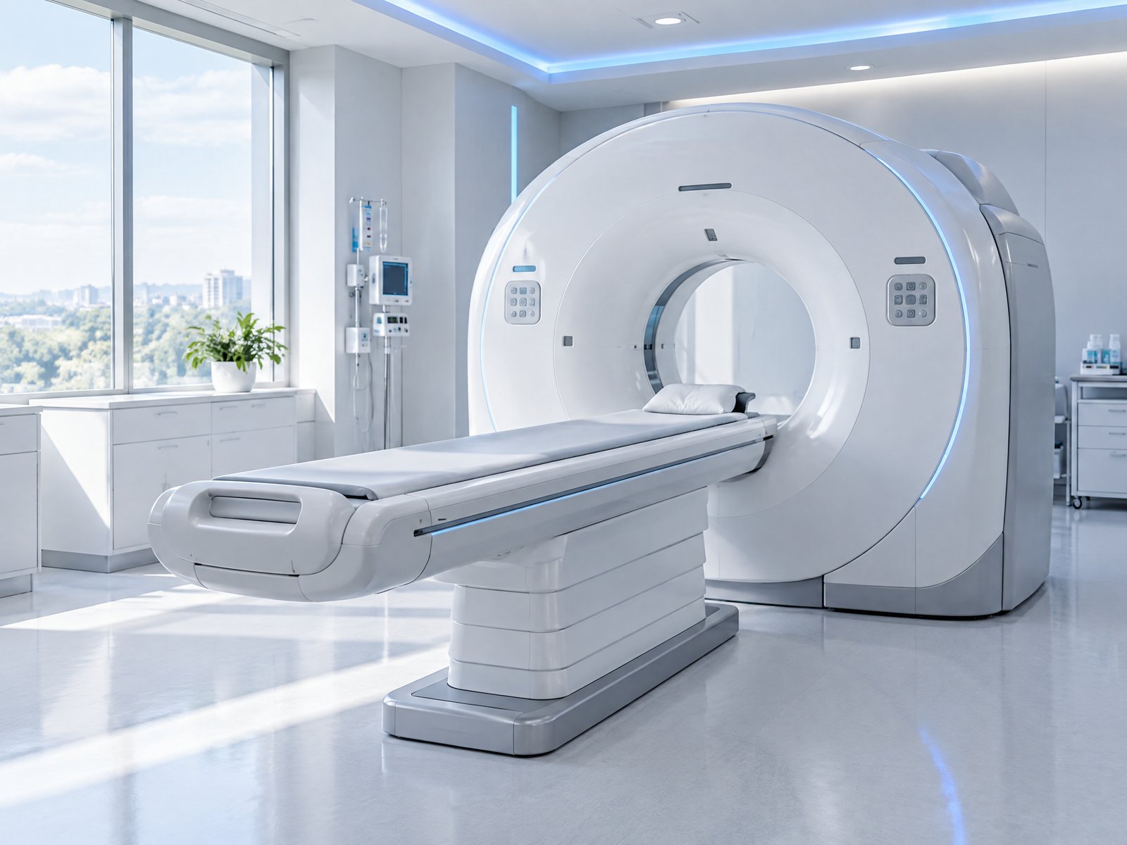
How deep learning is transforming the speed and accuracy of diagnostic imaging.
A single modern CT study can contain hundreds to thousands of image slices. A multi-sequence MRI exam can contain even more. Imaging volumes have grown steadily for years, while the number of radiologists has not kept pace.
The result is longer reporting backlogs, heavier workloads and greater risk of fatigue. Small, early-stage findings — the ones that matter most for outcomes — are the easiest to miss when a specialist is reading their hundredth study of the day.
Machine vision changes the equation. An AI model reviews every voxel of every slice with the same consistent attention, at any hour, in seconds.
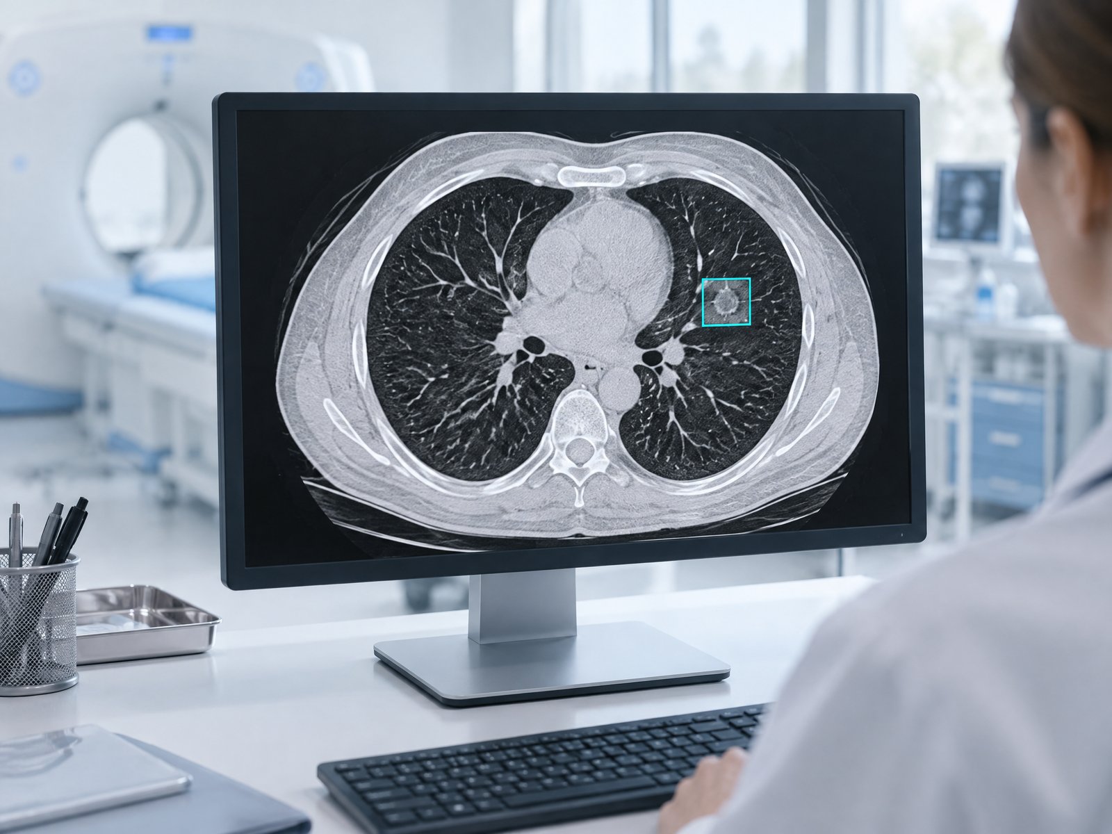
Machine vision uses deep neural networks — convolutional neural networks and, more recently, vision transformers — to learn visual patterns directly from data. Instead of being programmed with rules, the model is trained on large sets of expertly annotated scans until it learns what disease looks like at every stage.
Dinmar Labs' models work in three dimensions, analyzing the full scan volume rather than isolated 2D images. This lets them understand shape, texture, density and spatial relationships the way an experienced radiologist does — and pick up subtle signals spread across multiple slices.
Because AI models can be retrained as new data arrives, performance continues to improve over time.
Across the field, peer-reviewed research has shown that AI-assisted reading can match or exceed unaided human performance on specific detection tasks — and dramatically reduce the time it takes for urgent findings to reach a clinician.
| Measure | Traditional human read | Dinmar AI-assisted read | Impact |
|---|---|---|---|
| Time to first review of urgent finding | Hours to days (queue-dependent) | Minutes — auto-prioritized | Much faster |
| Analysis time per study | 10–30 minutes | Under 90 seconds (pre-read) | Much faster |
| Small nodule / early lesion detection | Variable; fatigue-sensitive | Consistent sensitivity* | Higher |
| Consistency across readers & shifts | Inter-reader variability | Identical, reproducible output | Higher |
| Longitudinal measurement | Manual, time-consuming | Automatic volumetric comparison | Automated |
| Performance at 3 a.m. | Fatigue risk | Unchanged | Reliable |
* Dinmar Labs internal retrospective validation and interim trial data. Results vary by indication and site. Dinmar Labs technology is investigational and intended to support, not replace, clinical judgment.
By pre-reading every study as it arrives and pushing critical suspected findings to the top of the worklist, AI turns a first-in-first-out queue into a priority queue. Patients with the most urgent conditions are seen first.
Acting as a second reader, AI catches findings that might otherwise be overlooked, while confidence scores help reduce unnecessary follow-ups from false positives.
Automating measurement and routine checks frees radiologists to spend their expertise where it matters most — complex cases and patient care.
Our CT models focus on early detection of lung nodules, liver and kidney lesions and other abnormalities, using Hounsfield-unit density analysis and 3D shape characterization to separate suspicious findings from benign structures.
For MRI, our models fuse information across multiple sequences (such as T1, T2 and FLAIR) to detect early neurological lesions and subtle tissue changes, with automatic quantification of volume and progression over time.
CT or MRI studies arrive securely from the scanner or PACS and are de-identified automatically.
3D machine-vision models examine the full volume, detecting and characterizing potential findings.
Suspected urgent findings are flagged and the study moves to the top of the radiologist's worklist.
Heatmaps, measurements and confidence scores are presented for clinician review and final diagnosis.
Request our research papers or apply to join the Early Adopter Program.
Request Papers Become an Early Adopter