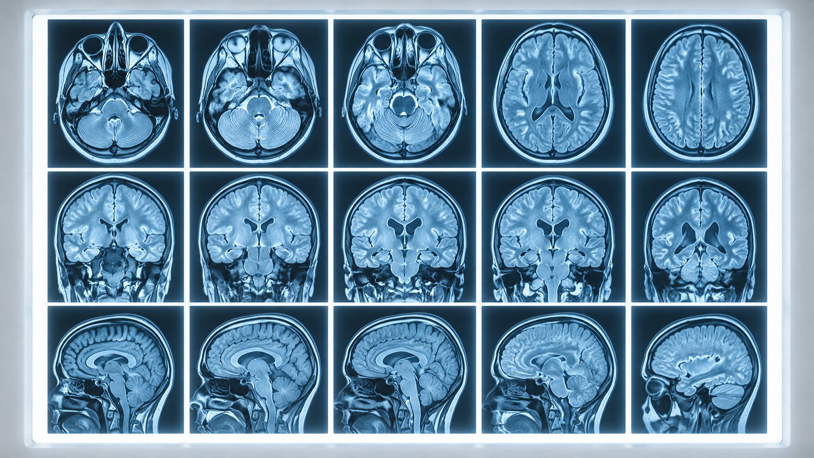
Our first models were built for CT. Today we're sharing a major technical step: Dinmar Labs' machine vision platform now analyzes brain MRI, with early results in detecting and quantifying neurological lesions.
MRI is a different challenge
CT measures tissue density on a standardized scale, which makes intensities fairly consistent from scanner to scanner. MRI is more complex. Its image intensities vary with the scanner, field strength and acquisition settings, and a single exam typically includes multiple sequences — such as T1-weighted, T2-weighted and FLAIR — each highlighting different tissue properties.
A lesion that is subtle on one sequence may be obvious on another. Radiologists mentally combine these views; our models needed to learn to do the same.
Our approach
- Intensity normalization to reduce scanner-to-scanner variation before analysis.
- Automatic co-registration so every sequence is precisely aligned voxel-to-voxel.
- Multi-channel 3D models that take all sequences as input simultaneously and learn which combinations of signals indicate disease.
- Volumetric quantification to measure lesion size and track change between exams.
Early results
In internal retrospective testing, the multi-sequence model detected small lesions with substantially higher consistency than our single-sequence baseline, and its automated volume measurements closely tracked expert manual segmentations. As always, these are early findings that will need independent validation.
A platform, not a single product
Extending from CT to MRI proves that our core technology — 3D machine vision, a trusted data pipeline and clinician-centered explainability — can be applied across modalities and diseases. That's central to our long-term vision.
Announcing Our First University Research Partnership
Opening Our Oxford Office